 ASVAB Camp
Learning goals and practical exam boundaries
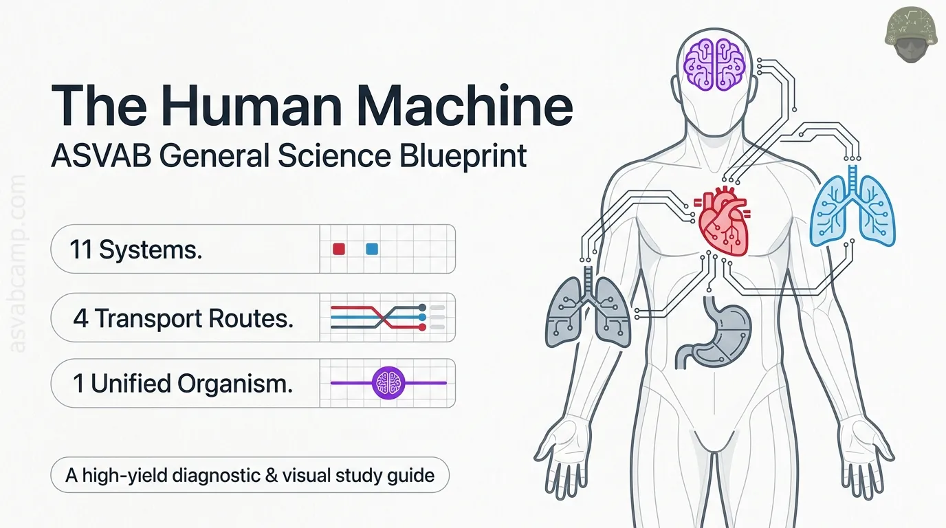
By the end of this lesson you should be able to name the eleven major organ systems and state the main job of each. You should be able to follow blood through the heart and lungs, food through the digestive tract, and urine from kidney to the outside. You should also be able to explain negative feedback, simple immune defenses, and how systems cooperate. The ASVAB tests broad functions and correct sequences, not rare diseases or fine anatomical detail.
Biological organization: tissues are not organs
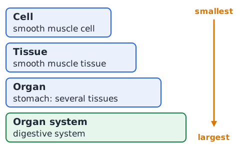
Living things are built in layers. Cells are the smallest living units. A tissue is a group of similar cells doing one shared job, such as muscle tissue or nervous tissue. An organ, such as the stomach or heart, is made of two or more tissue types working together. An organ system is a set of organs that cooperate on a larger task, and all the systems together make the organism. A common test trap is calling a tissue an organ, so check which level a question is asking about.
Homeostasis is dynamic regulation, not absolute stillness
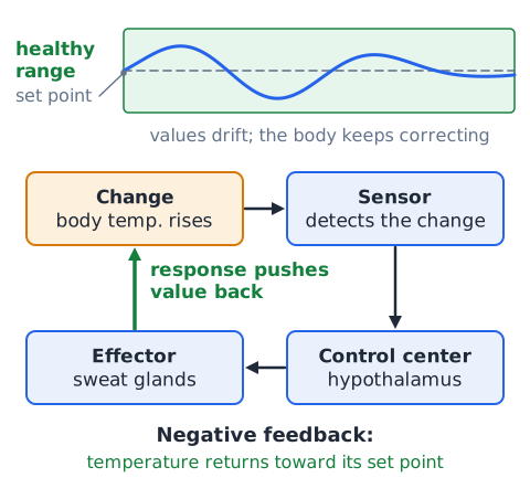
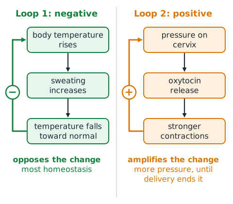
Homeostasis means keeping internal conditions, such as body temperature, blood glucose and water balance, within a healthy range. It does not mean nothing changes. Values drift up and down all day, and the body keeps correcting them. Most control works by negative feedback: a sensor detects a change, a control center responds, and an effector pushes the value back toward its set point. Positive feedback is rarer; it amplifies a change until some event ends it, as with contractions during childbirth.
Example 1 — trace system-level organization
Question: Arrange from smallest to largest: stomach, digestive system, smooth muscle cell, smooth muscle tissue. Step 1: The single cell is the smallest unit. Step 2: Many similar cells form smooth muscle tissue. Step 3: The stomach contains this tissue plus others, such as lining and connective tissue, so it is an organ. Step 4: The stomach works with other organs in the digestive system. Answer: cell, tissue, stomach, digestive system.
The eleven-system map
A useful map lists eleven systems. Integumentary: skin, hair and nails form a protective covering. Skeletal: bones support and protect. Muscular: muscles produce movement. Nervous: brain, spinal cord and nerves send fast signals. Endocrine: glands release hormones. Cardiovascular: heart and vessels move blood. Lymphatic and immune: return fluid and fight infection. Respiratory: lungs exchange gases. Digestive: break down food and absorb nutrients. Urinary: kidneys filter blood. Reproductive: make gametes and offspring.
Cardiovascular system: pump, vessels and circulating blood
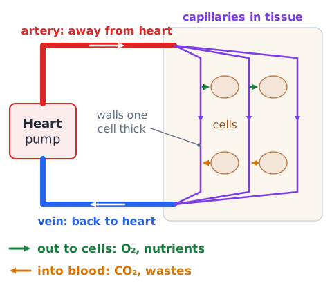
The cardiovascular system is the body's delivery network. The heart is its pump; its muscular walls contract rhythmically to push blood forward. Arteries carry blood away from the heart, veins bring it back, and tiny capillaries connect them inside the tissues. Blood carries oxygen, nutrients, hormones and heat out to cells and picks up carbon dioxide and other wastes. Because every cell depends on this delivery, a blocked vessel can starve tissue downstream even if the lungs work well.
Four heart chambers and one-way valves
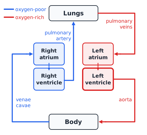
The human heart has four chambers. The two upper chambers, the atria, receive blood returning to the heart. The two lower chambers, the ventricles, pump blood out. The right side handles oxygen-poor blood from the body; the left side handles oxygen-rich blood from the lungs. The left ventricle has the thickest wall because it pushes blood through the whole body. Valves between chambers and at the exits act like one-way doors, closing to stop backflow, which produces the heartbeat sounds.
Pulmonary circulation and systemic circulation
Blood travels two loops. In the pulmonary loop, the right ventricle sends oxygen-poor blood through the pulmonary arteries to the lungs, where it gains oxygen and loses carbon dioxide; pulmonary veins return the now oxygen-rich blood to the left atrium. In the systemic loop, the left ventricle pumps blood into the aorta and out to the body, and the venae cavae return oxygen-poor blood to the right atrium. Remember: arteries are named for leaving the heart, not for oxygen content.
What red cells, white cells, platelets and plasma do
Blood has a liquid part and cells. Plasma, the pale yellow liquid that makes up just over half of blood, is mostly water and carries dissolved nutrients, salts, hormones, proteins and wastes. Red blood cells contain hemoglobin, an iron-containing protein that binds oxygen, so they are the main oxygen carriers. White blood cells defend against infection. Platelets are small cell fragments that help seal damaged vessels and start clotting. Most blood cells are produced in red bone marrow.
Capillaries and efficient exchange
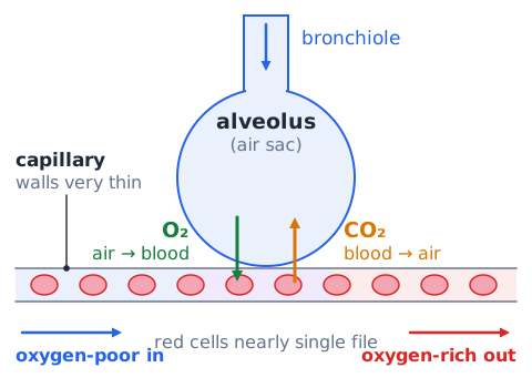
Capillaries are the smallest blood vessels, with walls only one cell thick. Red blood cells pass through them nearly single file, which keeps blood very close to surrounding cells. Exchange happens here by diffusion: oxygen and nutrients move out of the blood toward cells, while carbon dioxide and wastes move in. Diffusion works best across a thin barrier, over a large surface area, and with a strong concentration difference, which is why the body has billions of capillaries woven through its tissues.
Example 2 — solve an oxygen-poor blood route
Question: Oxygen-poor blood returns from the arm. Trace it to the lungs. Step 1: Systemic veins lead to the superior vena cava. Step 2: It enters the right atrium. Step 3: It passes a valve into the right ventricle. Step 4: The right ventricle pumps it into the pulmonary artery. Answer: vena cava, right atrium, right ventricle, pulmonary artery, lungs.
Respiratory system: airway path
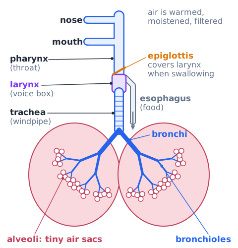
Air enters through the nose or mouth, where it is warmed, moistened and partly filtered. It passes through the pharynx, or throat, then the larynx, or voice box. The epiglottis, a flap of tissue, covers the larynx during swallowing so food goes down the esophagus instead. Air continues down the trachea, or windpipe, which splits into two bronchi, one for each lung. The bronchi branch into smaller bronchioles that end in clusters of tiny air sacs called alveoli.
Alveoli: where oxygen and carbon dioxide exchange
Alveoli are tiny, balloon-like air sacs wrapped in capillaries. They are where most oxygen enters the blood. Their walls and the capillary walls are extremely thin, and the lungs contain hundreds of millions of alveoli, giving a huge surface area. Oxygen diffuses from air into blood, and carbon dioxide diffuses from blood into air to be exhaled. Anything that thickens this barrier or destroys alveolar surface slows diffusion and lowers gas exchange.
The diaphragm and mechanics of breathing
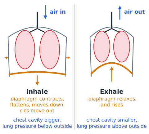
The diaphragm is a dome-shaped sheet of skeletal muscle beneath the lungs. When it contracts, it flattens and moves downward, and the rib muscles lift the ribs outward. This enlarges the chest cavity, lowering air pressure in the lungs below outside pressure, so air flows in. During quiet exhalation the diaphragm relaxes and rises, the chest cavity shrinks, pressure in the lungs rises, and air flows out. Air always moves from higher pressure to lower pressure; the lungs themselves have no muscle to pump.
Breathing versus cellular respiration
Breathing, or ventilation, is the physical movement of air into and out of the lungs. Cellular respiration is a chemical process inside each cell, mainly in the mitochondria, that uses oxygen to release energy from glucose and stores it as ATP. Carbon dioxide and water are its by-products. The two are linked: breathing supplies oxygen and removes carbon dioxide, and blood ferries both between lungs and cells. On the test, watch whether a question means air movement or energy release.
Example 3 — why does oxygen reach a leg muscle?
Question: A runner breathes hard. How does oxygen get from the air to a calf muscle? Step 1: Air reaches the alveoli. Step 2: Oxygen diffuses into pulmonary capillaries and binds hemoglobin in red cells. Step 3: Pulmonary veins return the blood to the left heart. Step 4: The left ventricle pumps it through the aorta and leg arteries. Step 5: In capillaries, oxygen diffuses into muscle cells. Answer: lungs and heart must both work; faster breathing cannot help if an artery is blocked.
Digestive system: breakdown, movement and absorption
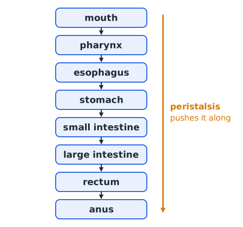
The digestive system turns food into molecules small enough to enter the blood. It does four broad jobs. Mechanical digestion physically breaks food apart by chewing and churning. Chemical digestion uses enzymes and acid to split large molecules such as starch, proteins and fats into smaller units. Movement, mostly by waves of smooth muscle contraction called peristalsis, pushes material along the tract. Absorption moves nutrients into the blood and lymph. Leftover material is then eliminated as feces.
Mouth, esophagus and stomach: distinct jobs
Each early section has its own role. In the mouth, teeth chew food and saliva moistens it; salivary amylase begins breaking down starch. The tongue shapes the food into a soft ball and pushes it back to be swallowed. The esophagus is a muscular tube that carries food from the throat to the stomach by peristalsis; it digests and absorbs essentially nothing. The stomach stores food, churns it, and adds acid and the enzyme pepsin, which begins protein digestion, producing a thick liquid called chyme.
Small intestine: the main site of nutrient absorption
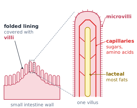
The small intestine is where most digestion is finished and most nutrients are absorbed. Here enzymes from the pancreas and the intestinal lining, helped by bile, complete the breakdown of carbohydrates, proteins and fats. Its inner surface is folded and covered with finger-like villi, which carry even smaller microvilli. These greatly increase surface area. Simple sugars and amino acids enter blood capillaries in the villi, while most digested fats enter small lymph vessels called lacteals.
Liver makes bile; gallbladder stores bile
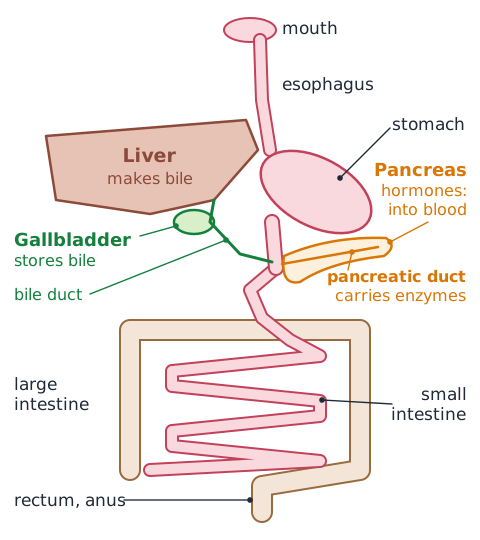
Do not mix up these two organs. The liver produces bile, a greenish fluid. The gallbladder, a small sac beneath the liver, only stores and concentrates bile, then releases it into the small intestine after a fatty meal. Bile is not an enzyme. It acts like a detergent, breaking large fat globules into tiny droplets so that fat-digesting enzymes, mainly pancreatic lipase, have more surface to work on. The liver also processes absorbed nutrients, stores glycogen, and breaks down toxins and old blood components.
Pancreas: one organ in two functional systems
The pancreas works in two systems. As a digestive organ, it sends pancreatic juice through a duct into the small intestine. This juice contains enzymes that digest carbohydrates, proteins and fats, plus bicarbonate that neutralizes stomach acid. As an endocrine gland, small clusters of cells called pancreatic islets release hormones into the blood. Insulin lowers blood glucose after a meal, and glucagon raises it when glucose falls. Read the question verb: secreting enzymes is digestive, releasing hormones is endocrine.
Large intestine and material exit
Material that is not absorbed in the small intestine passes into the large intestine. Its main jobs are to absorb much of the remaining water and some salts, which firms the waste into feces. Bacteria living there break down some leftover material and make certain vitamins, such as vitamin K. Feces are stored in the rectum until they leave through the anus. Feces are mostly undigested residue, bacteria and water, which is different from urine, the liquid waste the kidneys make by filtering blood.
Example 4 — classify an organ by the question verb
Question: Which system is described in this statement: the pancreas releases insulin into the blood? Step 1: Find the action, releases into the blood. Step 2: Identify the product, insulin, which is a hormone. Step 3: Hormones carried in the blood belong to the endocrine system. Answer: endocrine. If it said pancreatic enzymes flow into the intestine, the answer would be digestive.
Urinary system: filtration and fluid regulation
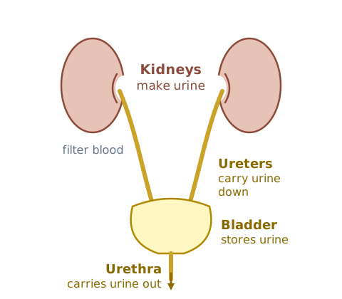
The urinary system cleans the blood and controls body fluids. The two kidneys filter blood, remove wastes such as urea, and make urine. In doing so they also regulate the amount of water in the body, salt levels, blood pH and, through fluid volume, blood pressure. Two ureters carry urine to the urinary bladder, a stretchy muscular sac that stores it. The urethra then carries urine out of the body. Only the kidneys make urine; the other parts move or hold it.
Nephron: glomerulus filters, tubules adjust
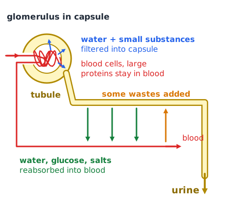
Each kidney contains about a million tiny filtering units called nephrons. A nephron begins with the glomerulus, a knot of capillaries inside a cup-shaped capsule. Blood pressure pushes water and small dissolved substances from the glomerulus into the capsule, while blood cells and large proteins stay behind. The filtrate then flows through a long tubule. Most water, glucose and useful salts are reabsorbed into the blood, and some wastes are added. What remains becomes urine.
Urinary transport sequence and common distractors
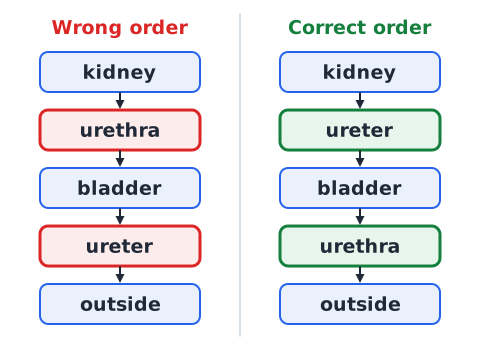
The correct route is kidney, then ureter, then bladder, then urethra, then outside. A memory aid: alphabetically, ureter comes before urethra, and so does its place in the route. The ureters run from kidney to bladder; the urethra leaves the bladder. Common distractors include swapping ureter and urethra, saying the bladder makes urine, or mixing in the gallbladder, which stores bile, not urine. The renal artery brings blood to the kidney; it does not carry urine.
Kidneys, hormones and homeostasis
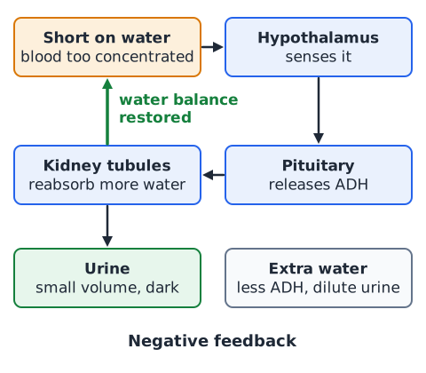
Kidneys respond to hormones to keep fluids balanced. When the body is short on water, the hypothalamus senses blood that is too concentrated and the pituitary releases antidiuretic hormone, ADH. ADH makes kidney tubules reabsorb more water, so urine becomes small in volume and dark. When there is extra water, less ADH is released and more dilute urine is produced. This is negative feedback. The kidneys also release a hormone that signals bone marrow to make more red blood cells when oxygen is low.
Example 5 — identify the urine transport mistake
Question: A diagram reads kidney, urethra, bladder, ureter, outside. Fix it. Step 1: The kidney makes urine, so it stays first. Step 2: The tube leaving each kidney is the ureter. Step 3: The bladder stores urine. Step 4: The urethra carries urine out. Answer: kidney, ureter, bladder, urethra, outside. The error was swapping ureter and urethra.
Nervous system: rapid information and coordination
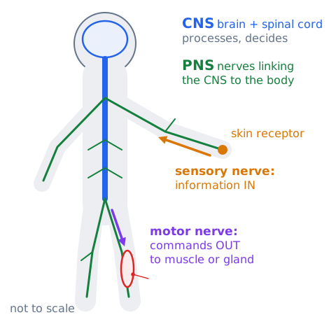
The nervous system carries fast electrical signals that let the body sense, decide and respond within fractions of a second. It has two main parts. The central nervous system, or CNS, is the brain and spinal cord; it processes information and makes decisions. The peripheral nervous system, or PNS, is the network of nerves that links the CNS to the rest of the body. Sensory nerves bring information in from receptors, and motor nerves carry commands out to muscles and glands.
Neuron structure and information direction
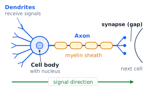
A neuron, or nerve cell, has three main parts. Dendrites are short branches that receive signals from other cells and carry them toward the cell body. The cell body holds the nucleus and keeps the cell alive. The axon is a long fiber that carries the signal away from the cell body toward the next cell. Many axons are wrapped in a fatty myelin sheath that speeds conduction. Signals cross a small gap called a synapse using chemical messengers called neurotransmitters.
Brain regions: focus on broad functions
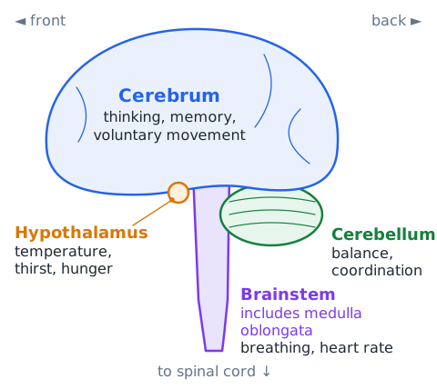
For the test, link each region to a broad job. The cerebrum is the largest part; it handles thinking, memory, language, voluntary movement and interpreting senses. The cerebellum, at the back beneath the cerebrum, coordinates smooth movement, posture and balance. The brainstem, which includes the medulla oblongata, connects to the spinal cord and controls automatic functions such as breathing rate and heart rate. The hypothalamus, below the cerebrum, regulates body temperature, thirst and hunger and links the nervous and endocrine systems.
Reflex arc: fast response pathways
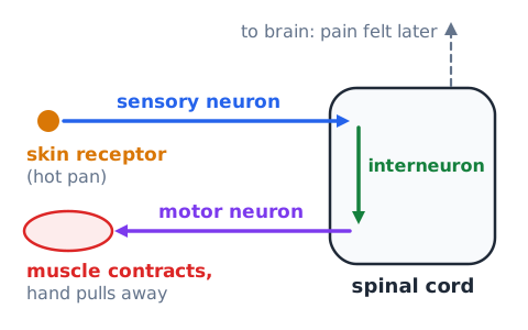
A reflex is an automatic, very fast response. In a withdrawal reflex, such as touching a hot pan, a sensory receptor in the skin sends a signal along a sensory neuron to the spinal cord. There, an interneuron passes it directly to a motor neuron, which makes a skeletal muscle contract and pull the hand away. Because the spinal cord handles the response without waiting for the brain, movement can start before you feel pain. The signal still travels up to the brain, which is why awareness follows a moment later.
Example 6 — locate the nervous structure for balance
Question: After an injury, a person walks unsteadily and has trouble with smooth, coordinated movement, but can still think clearly. Which brain region is most likely affected? Step 1: Thinking is a cerebrum job, and it is normal. Step 2: Balance and fine coordination are cerebellum jobs. Step 3: Breathing is not mentioned, so the brainstem is less likely. Answer: cerebellum.
Endocrine system: hormones in the blood
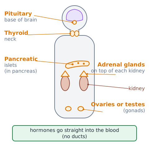
The endocrine system controls the body with chemical messengers called hormones. Endocrine glands, such as the pituitary, thyroid, adrenal glands, pancreatic islets, ovaries and testes, release hormones directly into the bloodstream rather than through ducts. Blood carries the hormone everywhere, but only target cells with matching receptors respond. Compared with nerve signals, hormone effects usually start more slowly and last longer, which suits jobs such as growth, metabolism, reproduction and blood glucose control.
Hypothalamus, pituitary and thyroid: signaling hierarchy
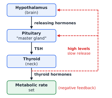
Many hormones work in a chain of command. The hypothalamus, part of the brain, monitors the body and sends releasing hormones to the pituitary gland just below it. The pituitary is often called the master gland because its hormones direct other glands. For example, it releases thyroid-stimulating hormone, TSH, which tells the thyroid in the neck to make thyroid hormones that set the body's metabolic rate. When thyroid hormone levels rise high enough, they signal the hypothalamus and pituitary to slow down, a classic negative feedback loop.
Pancreatic hormones and blood glucose
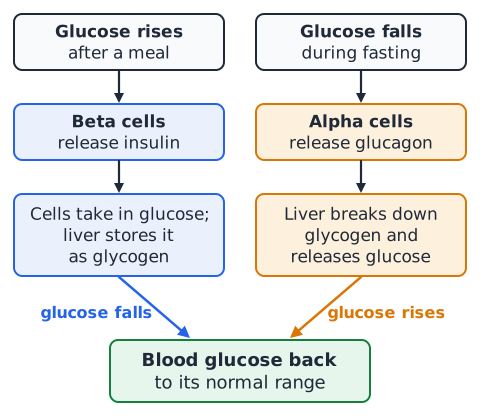
Scattered through the pancreas are small clusters of endocrine cells called pancreatic islets, or islets of Langerhans. They release two hormones with opposite effects on blood glucose. After a meal, when glucose rises, beta cells release insulin, which helps body cells take in glucose and signals the liver to store the extra as glycogen, so blood glucose falls. During fasting, when glucose drops, alpha cells release glucagon, which signals the liver to break down glycogen and release glucose, so blood glucose rises. Short version: insulin lowers, glucagon raises.
Adrenal glands and stress responses
One adrenal gland sits on top of each kidney, and each has two layers that behave like separate glands. The inner medulla, triggered by nerve signals, releases epinephrine (adrenaline) and norepinephrine within seconds of sudden stress. These raise heart rate, open the airways and free stored fuel for muscles, the fight-or-flight response. The outer cortex makes steroid hormones such as cortisol, which supports blood glucose during longer stress, and aldosterone, which helps the kidneys hold on to sodium and water.
Example 7 — positive versus negative feedback
Scenario: Loop 1: body temperature rises, sweating increases, and temperature falls back toward normal. Loop 2: during childbirth, pressure on the cervix triggers oxytocin release, which strengthens contractions, which raises the pressure further. Step 1: Ask whether the response opposes or amplifies the starting change. Step 2: Loop 1 opposes it, so it is negative feedback. Step 3: Loop 2 amplifies it until delivery ends the cycle, so it is positive feedback. Answer: Loop 1 negative, Loop 2 positive. Most homeostasis relies on negative feedback.
Skeletal system: support, protection, storage, blood cell origin
The adult skeleton has about 206 bones, along with cartilage and ligaments. It gives the body a supporting frame and anchors muscles so they can move body parts by working on bones as levers. It protects soft organs: the skull encloses the brain, the rib cage shields the heart and lungs, and the vertebrae surround the spinal cord. Bone stores minerals, mainly calcium and phosphorus, and releases them when blood levels fall. Red bone marrow makes red cells, white cells and platelets, a process called hematopoiesis.
Joints, cartilage, tendons and ligaments
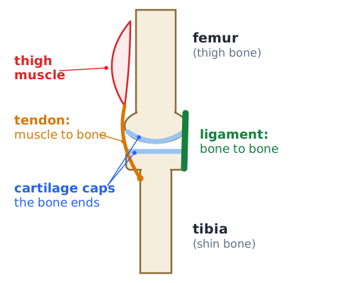
A joint, or articulation, is any place where two bones meet. Some joints, such as the sutures between skull bones, allow almost no movement, while others, such as the knee, elbow and shoulder, move freely. In freely movable joints the bone ends are capped with smooth cartilage that reduces friction and cushions impact. Ligaments are tough bands of connective tissue that connect bone to bone and keep a joint stable. Tendons are cords of connective tissue that connect muscle to bone and pass a muscle's pull to the skeleton.
Muscular system: three muscle types
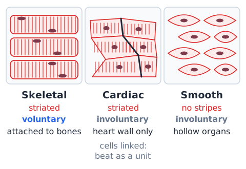
Skeletal muscle attaches to bones, looks striped (striated) under a microscope, and is voluntary, meaning you control it when you walk, lift or speak. Cardiac muscle is found only in the wall of the heart; it is also striated, but it is involuntary, and its cells are linked so a chamber contracts as a coordinated unit. Smooth muscle has no stripes and is involuntary; it forms the walls of hollow organs such as the stomach, intestines, bladder and blood vessels, where it squeezes contents along or changes vessel width.
Antagonistic muscle pairs and movement
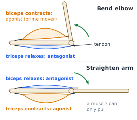
A muscle can only pull: it shortens when it contracts and cannot push. For that reason skeletal muscles usually work in opposing, or antagonistic, pairs across a joint. To bend the elbow, the biceps on the front of the upper arm contracts while the triceps on the back relaxes. To straighten the arm, the triceps contracts and the biceps relaxes. The muscle doing the main work is called the prime mover, or agonist; the one that relaxes and lengthens is the antagonist. The roles swap when the motion reverses.
Example 8 — distinguish tendon from ligament
Question: In a knee injury, a band joining the thigh bone to the shin bone is overstretched. Is it a tendon or a ligament? Step 1: Identify what the band connects: bone to bone. Step 2: Apply the rule: a ligament links bone to bone, while a tendon links muscle to bone. Answer: ligament. Memory aid: a tendon is the cord a muscle tugs on.
Integumentary system: skin, hair, nails and glands
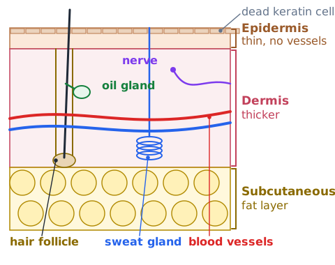
The integumentary system is the body's outer covering: skin, hair, nails, and the glands and sensory receptors in the skin. Skin is the largest organ. It blocks microbes, limits water loss and screens ultraviolet light; it helps regulate body temperature; it senses touch, pressure, pain and temperature; and it makes vitamin D in sunlight. Hair and nails are built mostly from the tough protein keratin. Sweat glands release watery sweat, and oil (sebaceous) glands release sebum that keeps skin and hair supple.
Epidermis, dermis and temperature control
Skin has two main layers resting on a fatty subcutaneous layer that insulates and stores energy. The epidermis is the thin outer layer; its surface cells are dead, flat and packed with keratin, and new cells keep moving up from below. It has no blood vessels. The dermis beneath is thicker and holds blood vessels, nerves, hair follicles and glands. When the body is hot, dermal vessels widen and sweat glands activate to shed heat; when it is cold, the vessels narrow to keep heat in.
Example 9 — why sweating can reduce temperature
Scenario: A soldier marches on a hot, dry afternoon and her body temperature starts to climb. Why does sweating help? Step 1: Sweat glands in the dermis release water onto the skin surface. Step 2: Changing liquid water into vapor takes energy, and much of that energy is drawn from the skin as heat. Step 3: Losing that heat cools the skin and the blood flowing just beneath it. Answer: evaporation carries heat away. Follow-up: in very humid air, sweat evaporates slowly, so cooling is weaker even if sweating continues.
Lymphatic system: fluid return and immune surveillance
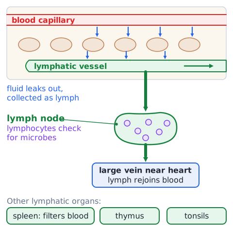
As blood passes through capillaries, some fluid leaks into the surrounding tissues. Lymphatic vessels collect this extra fluid, now called lymph, and return it to the bloodstream through large veins near the heart, which helps keep blood volume steady. Along the way, lymph flows through small lymph nodes, where white blood cells called lymphocytes check it for microbes and abnormal cells. Other lymphatic organs include the spleen, which filters blood, the thymus and the tonsils. Lymphatic vessels in the intestine also absorb dietary fat.
Innate defenses and adaptive responses
Innate defenses are present from birth, act quickly and respond the same general way to many kinds of invaders. They include physical barriers such as intact skin and mucous membranes, chemical barriers such as stomach acid, phagocytes that engulf microbes, and inflammation and fever. Adaptive defenses develop after exposure, take days to build and target one specific antigen. They depend on lymphocytes: B cells produce antibodies, and T cells direct the response or destroy infected cells. Adaptive immunity also leaves memory cells behind.
Antigen versus antibody and vaccines
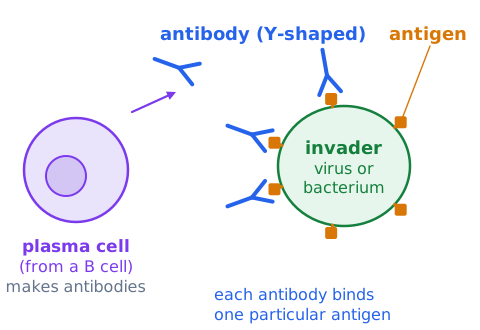
An antigen is any molecule, often a protein on the surface of a virus or bacterium, that the immune system recognizes as foreign. An antibody is a Y-shaped protein made by B cells, specifically by plasma cells derived from them, that binds to one particular antigen, blocking the invader or tagging it for destruction. A memory aid: the antigen generates the response, and the antibody is the body's answer. A vaccine presents a harmless form or piece of an antigen, so the body builds memory cells. A later encounter with the real germ then brings a faster, stronger response. That is active immunity; receiving ready-made antibodies is passive immunity.
Reproductive system: gametes and fertilization
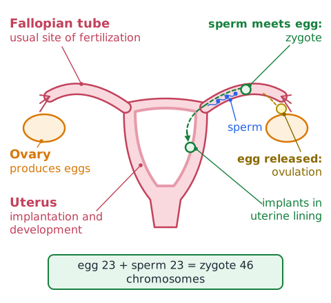
The reproductive system's job is producing offspring. The gonads make gametes, or sex cells: testes produce sperm and ovaries produce egg cells (ova). Gametes form by meiosis, so each carries a single set of 23 chromosomes instead of the usual 46. Gonads also make sex hormones: testosterone in the testes, and estrogen and progesterone in the ovaries. About once a month an ovary releases an egg, which is ovulation. Fertilization, the joining of sperm and egg, usually occurs in a fallopian tube and forms a zygote with 46 chromosomes.
Uterus, placenta and reproductive hormone roles
The uterus is a hollow, muscular organ where a fertilized egg implants and grows into an embryo and then a fetus. Its lining thickens each cycle under estrogen and progesterone; if no implantation occurs, the lining is shed during menstruation. After implantation, the placenta forms. It lets oxygen and nutrients pass from the mother's blood to the fetus and wastes pass back, without the two blood supplies mixing, and it makes hormones that maintain pregnancy. Near birth, oxytocin drives labor contractions.
Example 10 — identify production versus location
Question: Which organ produces egg cells, and which organ is where a fertilized egg develops? Step 1: Note the verb in each part: produce versus develop. Step 2: Gametes are produced in the gonads, so eggs come from the ovary. Step 3: After implantation, development takes place in the uterus. Answer: the ovary produces eggs; the uterus houses development. The fallopian tube is the usual place of fertilization, not of production.
Compare control systems: nervous versus endocrine
Both systems coordinate the body, but they differ in method and timing. The nervous system sends electrical impulses along neurons and chemical neurotransmitters across synapses. Its messages arrive in milliseconds, reach specific target cells and usually fade quickly, as when you jerk a hand away from a hot pan. The endocrine system releases hormones into the blood, which carries them everywhere, but only cells with matching receptors respond. Effects start more slowly and can last minutes to years, as with growth. The hypothalamus links the two systems.
Compare transport pathways: blood, air, food and urine
Each system moves material along a fixed route. Blood: body veins, right atrium, right ventricle, pulmonary artery, lungs, pulmonary vein, left atrium, left ventricle, aorta, body. Air: nose or mouth, pharynx, larynx, trachea, bronchi, bronchioles, alveoli. Food: mouth, pharynx, esophagus, stomach, small intestine, large intestine, rectum, anus. Urine: kidney, ureter, urinary bladder, urethra, outside. Knowing these four orders lets you spot a misplaced step quickly.
Compare protection: skin, skeleton, immune cells
Systems protect the body in different ways. Skin is a physical barrier: it keeps out microbes, slows water loss and absorbs some ultraviolet light. The skeleton is mechanical armor: the skull guards the brain, the rib cage guards the heart and lungs, and the vertebral column surrounds the spinal cord. Immune cells provide biological defense: phagocytes engulf invaders, and lymphocytes recognize specific antigens, and some of them develop into plasma cells that make antibodies. If a question is about blocking entry, think skin; about impact, think bone; about infection already inside, think immune cells.
Compare waste: CO2, urea and undigested residue
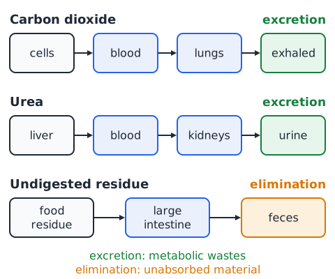
The body clears three different wastes by three routes. Carbon dioxide, made by cellular respiration in every cell, travels in the blood to the lungs and is exhaled. Urea, a nitrogen-containing waste formed in the liver when amino acids are broken down, is filtered from the blood by the kidneys and leaves in urine. Undigested food residue never entered body cells; it passes through the large intestine and leaves as feces. Excretion removes metabolic wastes, while elimination removes unabsorbed material.
Organ overlap is expected, not a contradiction
Many organs serve more than one system, so the same organ can appear under different headings. The pancreas releases digestive enzymes (digestive system) and insulin and glucagon (endocrine system). The pharynx carries both air and food. In males the urethra carries both urine and semen. Bones support the body and, through red marrow, also make blood cells. To classify an organ correctly, look at which function the question describes rather than where the organ sits.
Example 11 — solve a two-system exercise question
Question: During a hard run, why do both breathing rate and heart rate rise? Step 1: Working leg muscles speed up cellular respiration, using more oxygen and making more carbon dioxide. Step 2: Rising carbon dioxide in the blood is sensed by the brainstem, which signals the diaphragm and rib muscles to breathe faster and deeper, bringing more oxygen into the alveoli. Step 3: The heart beats faster and harder so the blood carries that oxygen to the muscles. Answer: the respiratory and cardiovascular systems cooperate.
Example 12 — separate nitrogen waste and food waste
Question: A student says urea and feces are both just leftover food. What is wrong? Step 1: Urea comes from breaking down amino acids inside the body; the liver turns toxic ammonia into urea, and the kidneys filter it into urine. Step 2: Feces are mostly undigested material, bacteria and water that never entered body cells. Answer: urea is a metabolic waste removed by the urinary system, while feces are eliminated by the digestive system.
Recognize common question stems efficiently
Many questions reuse a few stem patterns. Which organ produces X asks for a source, such as the liver for bile or the kidney for urine. Which structure carries X from A to B asks for a tube, such as the ureter, esophagus or pulmonary artery. Where does X mainly occur asks for a site, such as the alveoli for gas exchange or the small intestine for absorption. Which system includes X asks you to group organs. Identify the stem type first, then cross out choices from the wrong category before comparing what remains.
Precise vocabulary: paired distractors
Test writers like pairs of terms that sound alike or sit next to each other. The ureter carries urine from kidney to bladder; the urethra carries it out of the body. An artery carries blood away from the heart; a vein carries it back. A tendon joins muscle to bone; a ligament joins bone to bone. Insulin lowers blood glucose; glucagon raises it. An axon carries signals away from the cell body; dendrites bring signals in. The liver makes bile; the gallbladder stores it. Read every word.
Guided drill A — reconstruct cardiac flow
Practice: Put these in order for blood returning from the body: left ventricle, right atrium, pulmonary artery, lungs, left atrium, right ventricle, pulmonary vein, aorta. Answer: right atrium, right ventricle, pulmonary artery, lungs, pulmonary vein, left atrium, left ventricle, aorta. Check question: which chamber has the thickest muscular wall, and why? Answer: the left ventricle, because it pumps blood out to the entire body.
Guided drill B — trace three tubes
Practice: Name the tube for each trip. 1. Food from the throat to the stomach. 2. Air from the larynx toward the lungs. 3. Urine from a kidney to the bladder. Answers: 1. esophagus. 2. trachea, which then splits into the bronchi. 3. ureter. Bonus: which tube carries urine from the bladder to the outside? Answer: urethra. Note that the epiglottis covers the airway during swallowing.
Guided drill C — label systems by an action
Practice: Name the system that performs each action. 1. Filters blood and makes urine. 2. Releases hormones into the blood. 3. Exchanges oxygen and carbon dioxide with the air. 4. Returns tissue fluid to the blood. 5. Covers the body and produces sweat. Answers: 1. urinary. 2. endocrine. 3. respiratory. 4. lymphatic. 5. integumentary. Bonus: which system makes blood cells in red marrow? Skeletal.
Guided drill D — distinguish movement and production
Practice: Decide whether each organ mainly moves the substance or produces it. 1. Liver and bile. 2. Esophagus and food. 3. Kidney and urine. 4. Ureter and urine. 5. Ovary and egg cells. 6. Heart and blood. Answers: 1. produces. 2. moves. 3. produces. 4. moves. 5. produces. 6. moves; the heart pumps blood, while red bone marrow produces the blood cells.
Guided drill E — predict homeostatic feedback
Practice: Predict the corrective response. 1. Blood glucose climbs after a large meal. 2. Body temperature drops on a cold night. 3. Body water falls after heavy sweating. Answers: 1. The pancreas releases insulin, and glucose returns toward normal. 2. Skin blood vessels narrow and shivering produces heat. 3. More antidiuretic hormone is released, so the kidneys return more water to the blood and make a smaller volume of concentrated urine. All three are negative feedback.
Guided drill F — interdisciplinary problem
Practice: A hiker climbs to high altitude, where each breath holds fewer oxygen molecules. Predict three responses. Answers: 1. Respiratory: breathing becomes faster and deeper to bring in more oxygen. 2. Cardiovascular: heart rate rises at first to speed delivery. 3. Over days to weeks, the kidneys release the hormone erythropoietin, which signals red bone marrow to make more red blood cells. Bonus: which link here joins the urinary and skeletal systems? Answer: a kidney hormone acting on bone marrow.
Rapid recall — organ to function
Heart: pumps blood. Alveoli in the lungs: gas exchange. Diaphragm: main breathing muscle. Stomach: churns food and begins protein digestion. Small intestine: most digestion and nutrient absorption. Liver: makes bile and forms urea. Gallbladder: stores bile. Pancreas: digestive enzymes plus insulin and glucagon. Large intestine: absorbs water. Kidney: filters blood and makes urine. Bladder: stores urine. Brain and spinal cord: central nervous system. Cerebellum: balance and coordination. Skin: barrier and cooling. Red marrow: blood cells. Ovary: eggs.
Rapid recall — routes and exact contrasts
Routes: blood returning from the body goes right atrium, right ventricle, pulmonary artery, lungs, pulmonary vein, left atrium, left ventricle, aorta. Air goes trachea, bronchi, bronchioles, alveoli. Urine goes kidney, ureter, bladder, urethra. Contrasts: arteries leave the heart and veins return to it. Tendon is muscle to bone; ligament is bone to bone. Insulin lowers glucose; glucagon raises it. An antigen triggers a response; an antibody binds it. Negative feedback opposes change; positive feedback amplifies it.
How to study this lesson and take the quiz
Start with what each body system is made of and what it does. Next, follow the four main transport routes: air through the lungs, blood through the heart and vessels, food through the digestive tract, and waste through the kidneys. Finish with mixed questions that connect several systems.
The 40-question quiz explains every answer. The suggested time is our study recommendation, not an official ASVAB General Science time limit, and your quiz score is practice accuracy, not a prediction of your test result. After each attempt, go back to the questions you missed and say in your own words why each wrong choice looked tempting and why it fails.
 English ▾